From Surgery to Skin
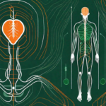
If you are on our website, you will very likely have heard something of the vagus nerve and how important it is to general health and well being. If you haven’t, you can look through our site or read some of our older blog posts to learn more about it.
Similarly, you may have heard of vagus nerve stimulation (VNS). Interest in VNS has grown with greater awareness of the nerve’s importance and there are now a range of techniques you can use. Some methods center around meditation and controlled breathing. Such techniques have roots in the ancient practices of meditation and yoga which are known to have significant health benefits.
Interest in electrical stimulation of the vagus nerve has grown in the last few decades. Initially, the medical application of this technique was both highly effective and highly invasive – involving major surgery to implant an electrical stimulator around the cervical (in the neck) branch of the vagus nerve.
Much more recently, interest has grown in non-invasive methods of VNS. This isn’t surprising, of course: invasive methods are expensive and carry considerable risks! All surgery is to some extent risky, but implanted VNS devices are also known to carry major side-effects: these include changes to tenor of a person’s speech and even complete loss of speech in some cases.
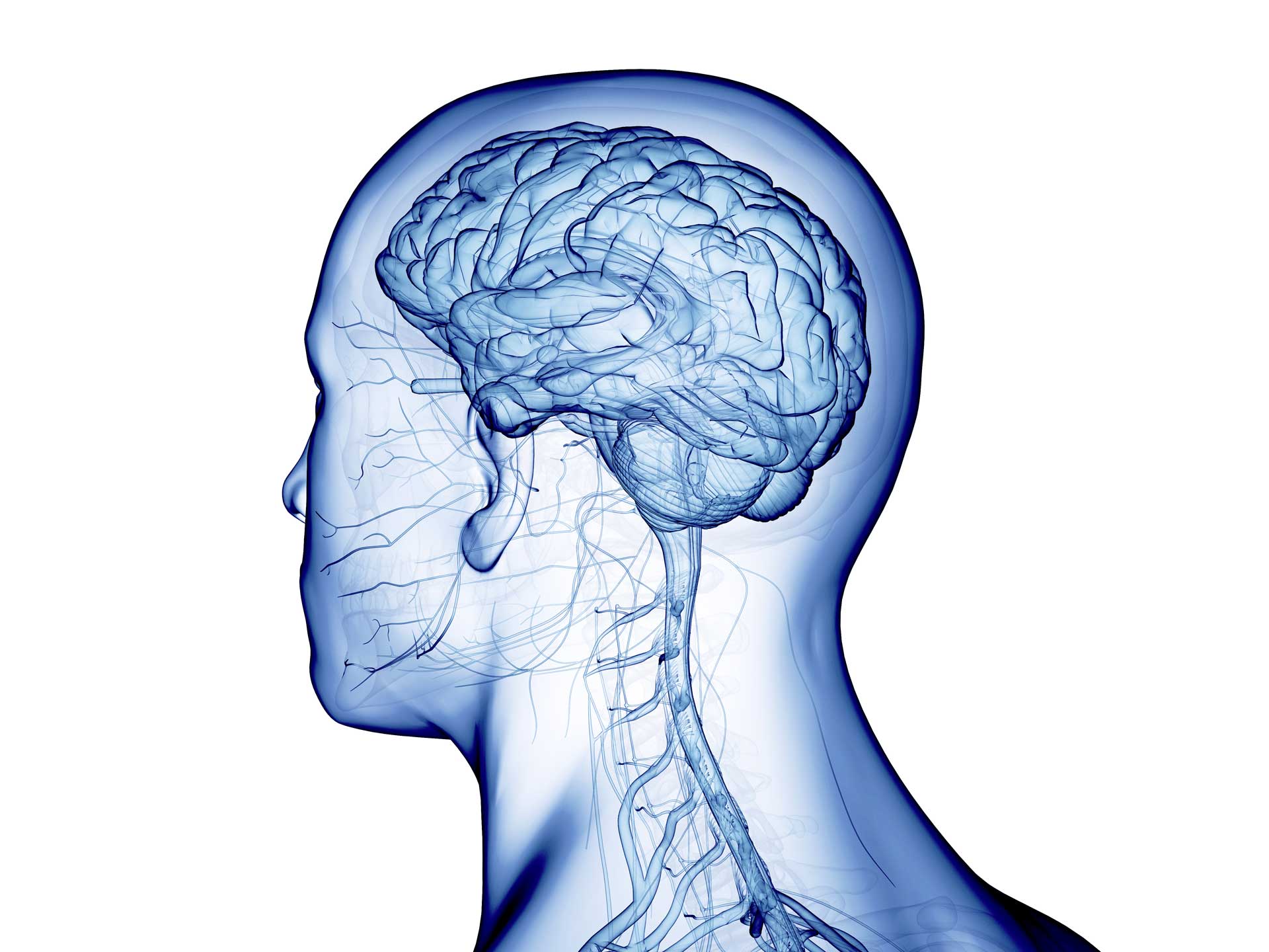
By contrast, much safer non-invasive methods of stimulating the cervical branch of the vagus nerve work by administering an electrical current from the surface of the skin that passes through a relatively thick layer of tissue (including skin, muscle and a tough arteria sheath), to stimulate the nerve. Electrodes are pressed against the neck, exactly where you would take your pulse and administered daily as when needed. This is obviously better than surgery, but scientists are still asking: are there even better ways to do this?
Safer and Easier
This has led scientists to increasingly become interested in stimulating the vagus nerve along it’s auricular branch – in the ear. At this location, the vagus nerve travels very close to the surface on the skin and has much less intervening tissue. What’s more, a growing body of research is demonstrating that there are very similar physiological responses to stimulation at either location and between invasive and non-invasive stimulation. Therefore, stimulating at the ear is becoming a very attractive option.
Targeting stimulation at the ear is actually anything but novel. Humans have been doing it for millenia. Think of Chinese medicine and its emphasis on acupuncture. When I first heard of acupuncture as an undergraduate science major I didn’t take it that seriously. I placed my trust firmly in modern science and medicine. But it turns out that the two are not mutually exclusive. Modern science is now providing strong empirical evidence for these ancient Eastern practices.
Knowledge of the passage of the nerves through the human body is usually obtained by the dissection of cadavers – bodies donated by people to the study of science. Over a few hundred years the art of dissection has steadily improved and with it the degree of intricacy of our knowledge of human anatomy.
Anatomical Knowledge
In 2002 two German medics dissected the ears of 7 cadavers (14 ears) to track the progress of innervation in the ear. This was an important event in the development of taVNS. It was the first attempt of its kind for almost a hundred years and remains the primary source of knowledge of the innervation of the ear. The study concluded that the cymba conchae and the tragus were the two primary locations that are innervated by the vagus nerve.
This year, in 2020, this anatomical knowledge was further extended by a group of Austrian scientists who used 3D episcopic imaging — a method in which repeated slices of tissue are photographed and then weaved together to create a 3D model — to further map the nerves and blood vessels in the human ear.
The image below is taken from the paper (click on it to view the original) which shows the kind of image produced by the scientific team.

The efficacy of this anatomical knowledge has since been tested extensively by scientists all over the world who have measured the effect of stimulating the auricular branch of the vagus nerve (ABVN) at both the tragus and the cymba conchae. These measures of physiological biomarkers can have either effects on brain function or bodily function.
The nerve fibres of ABVN are mainly afferent — which means they are essentially sensory and carry information back to the brain. The ABVN travels back from the ear to a region of the brainstem called the medulla oblongata. This is a junction point between signals deeper in the brain and further branches of the vagus nerve that travel throughout the body. It is via this junction point that stimulation of the ABVN can have an effect directly on pathways in the brain as well as pass signals down to the visceral organs.
This means that scientists can examine the effects of ABVN stimulation using biomarker directly from the brain and biomarker that may come from the body.
Brain Biomarkers
Neurological changes can be picked up by brain scans. Using such techniques, scientists can see that stimulation of the ABVN has very similar effects to both invasive and non-invasive stimulation of the vagus nerve at the cervical branch (in the neck). Below is an image from an fMRI study of tVNS to help sufferers of tinnitus. Again, you can click on the image to look at the original study.

Using such techniques, scientists have built up an understanding of how stimulation influences the brain. Explaining how to interpret these images and finding is out of the scope of this post, but what the study, and many like it, show is that targeting the auricular branch of the vagus nerve activates certain regions of the brain. The important point I’m trying to make here is that there is clearly an effect from tVNS that is measurable.
However, though many of these studies are of great interest to science and to the development of specific treatments using tVNS, one of the main reasons for using tVNS is it’s broader, more general health benefits.
Body Biomarkers
With this in mind, a better way of monitoring the effects of tVNS at home is by using two bodily biomarkers: heart rate variability (HRV) and blood pressure. Both these can be measured using equipment that is broadly available and relatively cheap to buy. They are also highly correlated to long term health outcomes.
Blood pressure has long been termed by doctors as the ‘silent killer’. Having a high blood pressure puts you at much greater risk of heart attacks and strokes; and greatly reduces quality of life in old age.
For a long time, heart rate was the other important measure for general health. But more recently, medics have realised that heart rate varies a lot in the short term — even between beats. And this heart rate variability (HRV) is a key measure of health with the same kind of predictive power as blood pressure. If you don’t know much about this, you can read our earlier post to get some background.
Both these measures are highly correlated with good vagus nerve health. So improving vagal tone (the strength of activity in the vagus nerve) is one of the best things you can do to improve your long term health outcomes.
Vagus nerve stimulation is not the only way of improving your blood pressure or HRV. In a previous post we covered a number of changes you can make to your lifestyle. But tVNS is one option which can help achieve a better balance.
As we’ve covered before, recent studies by UK scientists have indicated that a few weeks of tVNS can greatly improve HRV. And both these biomarkers can be measured at home using readily available equipment. You don’t have to carefully measure them to benefit from using tVNS, but if you want to check up on how you are responding to changes in your lifestyle and habits, these biomarkers will give you good feedback.
In upcoming posts we will explore how to go about this and what to expect.